The liver is an important organ in the human body that has many functions such as protein synthesis, drug biotransformation, and detoxification. Despite having strong regenerative abilities, severe liver damage can lead to liver failure. Liver transplantation, the main therapy for liver failure, still has many drawbacks such as difficulty in finding suitable donors, high costs, and long-term use of immunosuppressive drugs. These shortcomings have led to a high number of patient queues waiting for liver transplants. Many alternative therapies have been developed to address this problem, including hepatocyte transplantation, stem cell transplantation, Artificial Liver, and most recently, Bioartificial Liver (BAL). These alternative therapies still have many limitations in their application. All these limitations pose a challenge to reconstructing liver organoids that can be used for BAL materials, drug testing, and as models to understand the pathogenesis of liver disease.
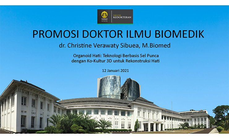
A doctoral candidate then carried out dissertation research from the FKUI Biomedical Science Doctoral Study Program, dr. Christine Verawaty Sibuea, M. Biomed. The research entitled “Liver Organoids: Stem Cell-Based Technology with 3D Co-Culture for Liver Reconstruction” was conducted to reconstruct the liver using stem cell-based liver organoids to regenerate the liver which can be used as a drug toxicity test model, as a model in studying disease mechanisms. liver, and as an ingredient for Bioartificial Liver (BAL). The liver is a complex unit consisting of parenchymal cells, namely hepatocytes and non-parenchymal cells.
Reconstruction of liver organoids requires cell components that replicate the in vivo microenvironment of the liver as well as culture techniques that can support liver function. Co-cultures of hepatocytes of hepatic stellate cell line (LX2), cord blood mesenchymal stem cells (UC-MSCs), and cord blood CD34+ stem cells (UCB-CD34+) are expected to present a microenvironment similar to the liver in vivo. The use of UC-MSCs in the co-culture of parenchymal and non-parenchymal cells provides a new opportunity to reconstruct liver organoids with a microenvironment that mimics the liver in vivo and has a liver function. Liver organoids in this study were reconstructed from hepatocytes, hepatic stellate cells (LX2), umbilical cord mesenchymal stem cells (UC-MSCs), and umbilical cord blood hematopoietic stem cells (UCB-CD34+). Mouse primary hepatocytes, LX2, UC-MSCs, and UCB-CD34+ were co-cultured in 11 ratio formulations and in 4 types of culture medium to obtain optimal ratio and culture medium. Hepatocyte ratio: LX2: UC-MSCs: UCB-CD34+ with a ratio of 5:1:2:2 which is the optimal ratio, cultured in Williams E optimal culture medium supplemented with PRP, ITS, and dexamethasone for 14 days and analyzed for morphology, function liver, and angiogenic potential. The findings in this study showed that the viability of liver organoids could last up to day 14 and that the viability of liver organoids was much better than monoculture. The morphology of organoids is solid and bulging with cell nuclei on the surface and partly inside the organoid. Albumin protein expression, GOT protein expression, and liver organoid CD31 protein expression was stable until day 14 and much better than monoculture. Liver organoid albumin gene expression increased until day 14, while GOT gene expression decreased until day 14. Hepatic organoid urea secretion decreased until day 5 and albumin secretion decreased until day 7.
In conclusion, studies of liver organoids reconstructed from primary hepatocytes, LX2, UC-MSCs, UCB-CD34+ with an optimal ratio of 5:1:2:2 in simple and economical Williams E optimal culture medium supplemented with PRP, ITS, and dexamethasone, were able to maintain viability and function up today 14. Liver organoids in this study can be used as models for drug testing and can be developed to become LAB materials. The results of the study were presented by dr. Christine Verawaty Sibuea, M. Biomed in her doctoral promotion session which took place virtually on Tuesday, January 12, 2021, at 10.00 WIB. With straightforward dr. Christine managed to defend her dissertation by answering questions and objections from the examiner team, chaired by Prof. Dr. dr. Rino A. Gani, SpPD-KGEH with members of the testing team Prof. Dr. dr. Sri Widia A. Jusman, MS; dr. Imelda Rosalyn Sianipar, M. Biomed., Ph.D.; and dr. Hannah. M. Kes., PhD., AiFO from Padjajaran University. The promoter in this research is Prof. dr. Jeanne A. Pawitan, MS, Ph.D. with co-promoter dr. Radiana Dhewayani Anatrianto, M. Biomed, PhD and dr. Chyntia Olivia Maurine Jasirwan, PhD., SpPD-KGEH. At the end of the session, the chairman of the session who is also the Dean of FKUI Prof. Dr. dr. Ari Fahrial Syam, SpPD-KGEH, MMB, appointed dr. Christine Verawaty Sibuea, The liver is an important organ in the human body that has many functions such as protein synthesis, drug biotransformation, and detoxification. Despite having strong regenerative abilities, severe liver damage can lead to liver failure. Liver transplantation, the main therapy for liver failure, still has many drawbacks such as difficulty in finding suitable donors, high costs, and long-term use of immunosuppressive drugs.
These shortcomings have led to a high number of patient queues waiting for liver transplants. Many alternative therapies have been developed to address this problem, including hepatocyte transplantation, stem cell transplantation, Artificial Liver, and most recently, Bioartificial Liver (BAL). These alternative therapies still have many limitations in their application. All these limitations pose a challenge to reconstructing liver organoids that can be used for BAL materials, drug testing, and as models to understand the pathogenesis of liver disease. A doctoral candidate then carried out dissertation research from the FKUI Biomedical Science Doctoral Study Program, dr. Christine Verawaty Sibuea, M. Biomed. The research entitled “Liver Organoids: Stem Cell-Based Technology with 3D Co-Culture for Liver Reconstruction” was conducted to reconstruct the liver using stem cell-based liver organoids to regenerate the liver which can be used as a drug toxicity test model, as a model in studying disease mechanisms. liver, and as an ingredient for Bioartificial Liver (BAL). The liver is a complex unit consisting of parenchymal cells, namely hepatocytes and non-parenchymal cells.
Reconstruction of liver organoids requires cell components that replicate the in vivo microenvironment of the liver as well as culture techniques that can support liver function. Co-cultures of hepatocytes of hepatic stellate cell line (LX2), cord blood mesenchymal stem cells (UC-MSCs), and cord blood CD34+ stem cells (UCB-CD34+) are expected to present a microenvironment similar to the liver in vivo. The use of UC-MSCs in the co-culture of parenchymal and non-parenchymal cells provides a new opportunity to reconstruct liver organoids with a microenvironment that mimics the liver in vivo and has a liver function. Liver organoids in this study were reconstructed from hepatocytes, hepatic stellate cells (LX2), umbilical cord mesenchymal stem cells (UC-MSCs), and umbilical cord blood hematopoietic stem cells (UCB-CD34+). Mouse primary hepatocytes, LX2, UC-MSCs, and UCB-CD34+ were co-cultured in 11 ratio formulations and in 4 types of culture medium to obtain optimal ratio and culture medium. Hepatocyte ratio: LX2: UC-MSCs: UCB-CD34+ with a ratio of 5:1:2:2 which is the optimal ratio, cultured in Williams E optimal culture medium supplemented with PRP, ITS, and dexamethasone for 14 days and analyzed for morphology, function liver, and angiogenic potential. The findings in this study showed that the viability of liver organoids could last up to day 14 and that the viability of liver organoids was much better than monoculture.
The morphology of organoids is solid and bulging with cell nuclei on the surface and partly inside the organoid. Albumin protein expression, GOT protein expression, and liver organoid CD31 protein expression was stable until day 14 and much better than monoculture. Liver organoid albumin gene expression increased until day 14, while GOT gene expression decreased until day 14. Hepatic organoid urea secretion decreased until day 5 and albumin secretion decreased until day 7. In conclusion, studies of liver organoids reconstructed from primary hepatocytes, LX2, UC-MSCs, UCB-CD34+ with an optimal ratio of 5:1:2:2 in simple and economical Williams E optimal culture medium supplemented with PRP, ITS, and dexamethasone, were able to maintain viability and function up today 14. Liver organoids in this study can be used as models for drug testing and can be developed to become LAB materials. The results of the study were presented by dr. Christine Verawaty Sibuea, M. Biomed in her doctoral promotion session which took place virtually on Tuesday, January 12, 2021, at 10.00 WIB. With straightforward dr. Christine managed to defend her dissertation by answering questions and objections from the examiner team, chaired by Prof. Dr. dr. Rino A. Gani, SpPD-KGEH with members of the testing team Prof. Dr. dr. Sri Widia A. Jusman, MS; dr. Imelda Rosalyn Sianipar, M. Biomed., Ph.D.; and dr. Hannah. M. Kes., PhD., AiFO from Padjajaran University. The promoter in this research is Prof. dr. Jeanne A. Pawitan, MS, Ph.D. with co-promoter dr. Radiana Dhewayani Anatrianto, M. Biomed, PhD and dr. Chyntia Olivia Maurine Jasirwan, PhD., SpPD-KGEH. At the end of the session, the chairman of the session who is also the Dean of FKUI Prof. Dr. dr. Ari Fahrial Syam, SpPD-KGEH, MMB, appointed dr. Christine Verawaty Sibuea,M.Biomed became a Doctor in the Doctoral Program in Biomedical Sciences at FKUI. Through his remarks, the Dean said, “I hope that Dr. dr. Christine will continue this research. Because earlier what he did was the first in Indonesia to carry out this reconstruction. We will even patent some of the techniques later because this is the first in the world.” (Source of FKUI Public Relations: https://fk.ui.ac.id/berita/peneliti-fkui-kembangkan-technology-based-sel-punca-dengan-ko-kultur-3d-untuk-reconstruction-hati.html)

