The heart is a vital organ responsible for pumping blood throughout the body. If a person is born with abnormalities in the heart’s structure, such as congenital heart disease (CHD), the heart may have difficulty performing its function. Therefore, various studies continue to be carried out to find new therapies that can help repair heart tissue in CHD patients.
Building on that challenge, a doctoral student in the Biomedical Sciences Doctoral Program (PDIB), Faculty of Medicine, Universitas Indonesia (FKUI), Ariyani Noviantari,conducted a study entitled: “The Effect of Polyvinyl Alcohol – human Fibroblast-Derived Matrix (PVA/hFDM) Hybrid Scaffold on the Differentiation of Human Umbilical Cord Mesenchymal Stem Cells toward the Cardiomyoblast Lineage In Vitro”. This study used mesenchymal stem cells derived from the human umbilical cord, which were then directed to develop into heart muscle cells, or cardiomyocytes. The study also used a special material called a scaffold as a growth
site for the cells, in the hope of helping the cells develop into heart muscle cells more effectively.
This research is part of the development of regenerative medicine, an approach that aims to help the body repair damaged tissue. The use of stem cells combined with a scaffold is expected to become one approach to helping the process of repairing heart tissue in the future, so that it has the potential to be developed in research on congenital heart disease.
Stem cells can be likened to basic cells that have the ability to develop or differentiate when given certain conditions. In this study, stem cells from the umbilical cord were given special conditions so that they would develop into cells with characteristics similar to heart muscle cells. This process was then observed to determine what occurs during the differentiation process.
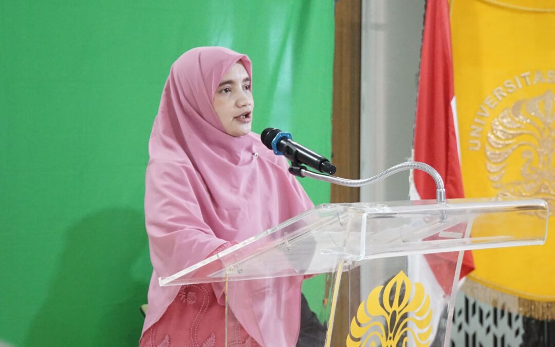
In the series of the PDIB FKUI Doctoral Promotion, Ariyani explained that cells need an environment in which to grow. To provide such a growth site for the cells, this study used polyvinyl alcohol – human fibroblast-derived matrix, or what is called the PVA/hFDM scaffold. This material can be imagined as a house or framework where
cells grow and develop.
PVA is an artificial or synthetic material in gel form, while hFDM contains substances derived from the natural environment of human fibroblast cells. The two are combined to create a three-dimensional material that is expected to provide a place that can support cell growth. The researcher then compared cells grown on a flat surface, such as in a laboratory culture dish, with cells grown on the three-dimensional PVA/hFDM scaffold.
The results of the study showed an interesting finding: cells grown in a three-dimensional environment did not do as well as cells grown on a flat surface. Some markers indicating the change of cells into heart muscle cells were in fact found at higher levels in cells grown on a flat surface. In addition, the cellular changes that occurred in this study were still at an early stage, with characteristics of heart muscle cells or cardiomyocytes that were not yet mature – often called cardiomyoblasts – and had not developed into fully mature heart muscle cells.
Ariyani, a researcher at the Center for Biomedical Research, Health Research Organization, National Research and Innovation Agency (BRIN), explained that these findings provide an important lesson for the development of regenerative medicine technology. Until now, a three-dimensional environment has been considered capable of providing conditions closer to those inside the body. However, this study shows that a three-dimensional environment alone is not enough to make cells develop into heart muscle cells more effectively. Many other factors can influence cell development, such as the material used as the growth site, the stiffness of the material, how cells attach to the material, and the environmental conditions during cell development. Therefore, this serves as a basis for researchers to continue refining the materials and conditions that are most suitable so that cells can develop into heart muscle cells more effectively.
Ariyani stated that the results of this study also cannot yet be used directly as a treatment for patients. This research is still at the laboratory stage and requires various subsequent research stages to ensure its safety and benefits. Further research will investigate more deeply whether cells that have been directed to become heart muscle cells truly have the ability to work like normal heart muscle cells. Researchers also need to know whether the cells can grow and develop well, and interact with the surrounding tissue. The PVA/hFDM scaffold that has been developed is also expected to continue to be optimized, so that in subsequent
studies it can provide a more suitable environment to support the growth and development of stem cells into more mature heart muscle cells.
In closing, Ariyani expressed her hope that this research can become a reference for scaffold development in heart tissue engineering, for example as a cell model for the treatment of congenital heart disease. The results of this study are also expected to become preliminary data on the use of the PVA/hFDM scaffold for the differentiation of mesenchymal stem cells into heart muscle cells, so that they can contribute to the advancement of biomedical science and open new opportunities in heart tissue repair in the future.